John Stuart Douglas PhD
“I was just 9 years old when my mother gave me a small microscope which magnified 100 times. The first time I observed a drop of water from our birdbath, I saw a curious microscopic creature which was shaped like a bell on a long stalk. Suddenly, the bell was gone! Then miraculously it slowly reappeared as the stalk uncoiled. The Vorticella started to feed using a ring of beating cilia creating a current bringing food to its mouth”
My lifelong adventure and love of microscopy had begun.
In August 2025, 74 years later with a PhD in Marine Microbiology, I still am enthralled by watching and learning from the Vorticellids. Antonie van Leeuwenhoek was the first person to observe Vorticella through a microscope, describing it in a letter to the Royal Society in 1676. Antonie called it the “Bell Animalcule” relating to its cell shape. Antonie used a microscope he made with a single lens magnifying 100 times.
Introduction
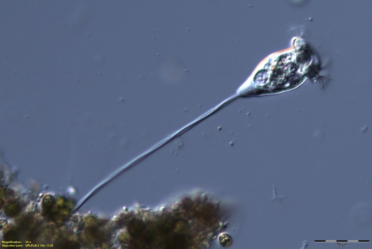
In this paper I will be concentrating on the two elements of vorticella which defines them: The adoral feeding cilia membranella, and the contractile stalk.
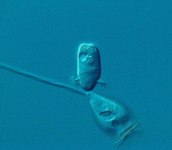
The genus Vorticella is characterized by bell-shaped bodies with a large oral area surrounded by cilia and a stalk for attachment to a substrate.
The genus Vorticella has many species, the number of which varies according to the author. For example: R. Kudo 5th Ed 1977 cites 5 species, H. Strebel 2001 cites 4 species, A. Kahl 1931 cites over 70 species. Whilst in the literature over 200 species of Vorticella have been described, many may be synonyms. Modern phylogenetic research places many of the 200 into other peritrichs groups and clades.
Feeding:
I have the luxury of owning an Olympus BX53 upright research microscope with led illumination, DIC, phase contrast, brightfield, and darkfield modes. All managed by advanced Olympus Dimension software.
A recently added SC 180 Olympus camera (replacing the DP 28) allows very high resolution, very high shutter speeds along with 18.1 million small 1.25-micron pixels.
To understand the world of microbes we must delve into fluid dynamics where the Reynolds Number equation dominates: [Re = (ρ * v * L) / μ].
We large animals never think about what would happen if we were to shrink to the size of a microbe. At the scale of protozoans and other microorganisms, viscous forces are dominant, meaning the fluid behaves as if it were more “sticky”. This leads us to laminar flow, where fluid moves in smooth, parallel layers, making it easier for microbes to attach to surfaces like pipes or other structures.
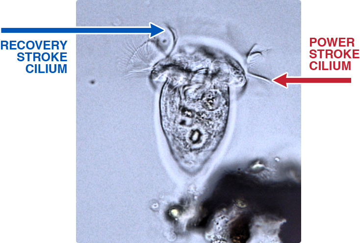
How does this apply to the case of Vorticella, where the adoral ring of cilia appear to move with amazing speed, just a blur looking under the microscope? However, the SC180 camera can stop them in their tracks enabling their motion to be analysed (fig 2).
So how do they propel themselves through this “molasses” as fast as we see them under the microscope?
Vorticella’s cilia move like miniature oars with a difference, unlike oars they are flexible. During the power stroke the cilia are stiff and fully extended creating a lot of drag; but during the recovery stroke the cilia flex and curl, creating significantly less drag. The power stroke creates a vortex in the surrounding fluid, bringing food to the vorticellid.
That is why humans rowing stiff oars must lift their oars out of the water on the recovery stroke.
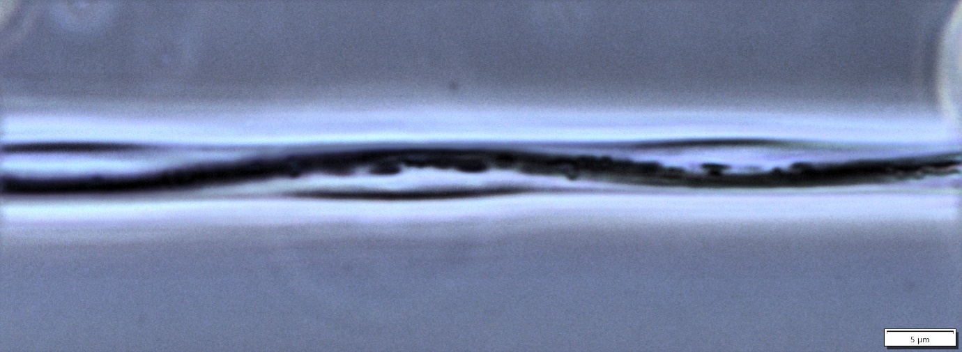
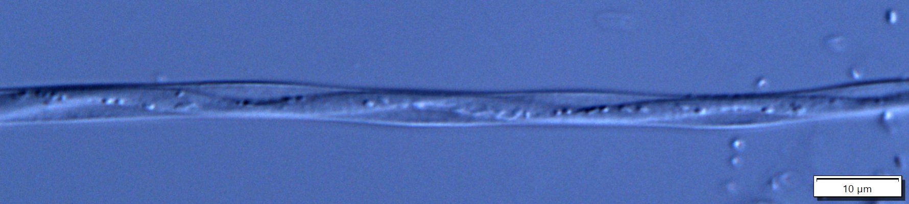
The Contractile Stalk:
The vorticella stalk contracts with an amazing speed taking only 1/60 of a second to fully contract. It is thought that contraction results from the entropic collapse of spasmin via Ca2+ screening of electrostatic repulsion. The released Ca2+ ions bind to a 20-kDa calcium-binding protein called spasmin, which constitutes 40–60% of the spasmoneme dry mass. As a result, a state of tension is induced in the spasmoneme which drives its contraction with a maximum speed of ∼6 cm s−1 (Mechanics of Vorticella Contraction, Gaurav Misra et al; Biophys J. 2010 Jun 16;98(12):2923–2932).